VERTEBROPLASTY
Indications
Vertebral compression fractures
Painful vertebral Hemangiomas
Vertebral metastasis
Multiple Myeloma with vertebral involvement
Procedure
Vertebroplasty is a minimally invasive procedure that provides immediate relief , continued and prolonged relief that may increase the patient's daily activity level, which in turn helps provide a better quality of life.
Vertebroplasty is performed to provide pain relief and to produce bone strengthening and vertebral stabilization when the lesion threatens the stability of the spine. Radiation therapy may be performed in conjunction with vertebroplasty. Radiation therapy does not interfere with the mechanical properties of bone cement and complements.
Vertebral fractures are the most common complication of osteoporosis. Age-related osteoporotic compression fractures occur in more than 500,000 patients per year in the United States. About 15% of women in the United States older than 50 years of age will suffer one or more vertebra compression fractures related to osteoporosis. Patients most likely to benefit from vertebroplasty are those with a focal, intense, deep pain associated with evidence of a new or progressive compression fracture. Patients who seem to respond the best include those with a single level or a few levels for treatment, fractures that are present less than 2 months or a recent worsening of the fracture, and no significant sclerosis of the fractured vertebra. It should also be determined if the patient is able to tolerate lying prone for 1 to 2 hours.
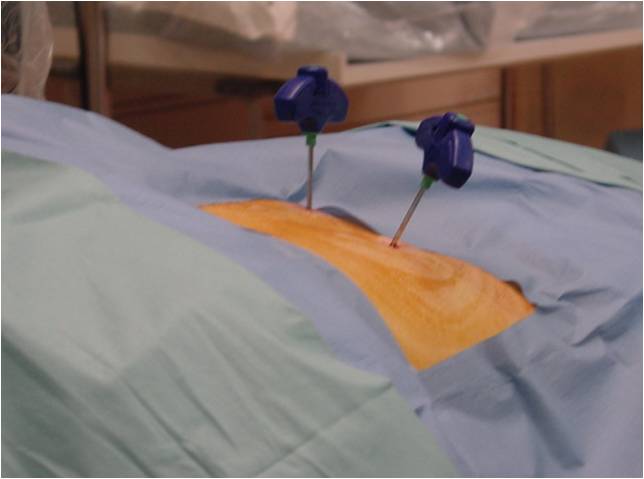
The procedure is done under conscious sedation. Sedative analgesics like fentanyl, midazolam, or propofol are administered. Prior to the start of the procedure, a prophylactic antibiotic should be given intravenously. The patient is taken to the fluoroscopy suite and placed in the prone position on the operating room table with all pressure points padded and standard monitoring applied. Care is taken to add extra padding to the table due to the osteoporotic spine and risk of rib fractures.
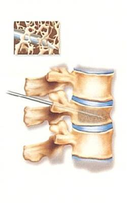
The level for the procedure is located in the posteroanterior fluoroscopic view. The fluoroscope is then rotated to an oblique posi�tion to maximize the oval appearance of the pedicle. Proper view is identified by seeing a "Scottie dog" appearance of the pedicle. It is important to "flatten out" the end plates of the vertebral bodies; if necessary, cephalocaudad or caudocephalad fluoroscopic angle may improve the image. This will locate the pedicle well within the vertebral body in the fluoroscopic view. This facilitates a central placement of the needle. The target for entry is the superior lateral quadrant of the pedicle in the fluoroscopic view.
After the site of entry is identified, a skin is anaesthetized. A small skin incision is made with a No. 11 blade scalpel to allow insertion of a large bore biopsy needle. An 11 gauge bone biopsy needle is inserted through the incision. It is advanced in "gun-barrel" fashion in the oblique fluoroscopic view to monitor direction. The oblique view will show the needle shaft end-on as a circle within the center of the pedicle. Once contact has been made with bone, insertion through the cortex of the bone may be accomplished with a twisting motion of the needle or gentle tapping of the needle with a sterile hammer. It is important to "set" the needlepoint at the exact site of entry.
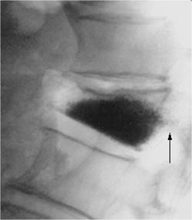
In the lateral view, the needle is advanced to the junction of the anterior and middle third of the vertebral body. After placement of the needle, the cement is prepared for injection. The amount to be mixed is based on the clinical observation of an average of 7 to 8 mL of cement injected in a lumbar vertebral body. The liquid monomer is titrated until it takes on toothpaste like consistency. There are several delivery systems available that may be used to simplify the mixing and delivery in addition to decreasing fumes. If these systems are not available, 2-mL syringes may be filled and used one at a time to inject the cement into the vertebral body. The injection of the cement is followed under fluoroscopic guidance in the lateral view. When the spread of the cement starts to invade the posterior one third of the vertebral body, injection should be stopped. After the procedure, the patient should be monitored for 3 hours in a recovery area prior to dismissal. The patient should be supine for 2 hours and then may sit. A CT image through the levels (2-mm slices) is recommended for documentation purposes. The patient is dismissed with routine pain medications and a graduated resumption of activity. Discharge instruction for the patient should include advice to call the physician for the following symptoms: New onset of back pain, Chest pain, Lower extremity weakness, Fever > 100 �F.
Contraindications of vertebroplasty are epidural involvement of the infiltrative lesion, Coagulation disorders, extensive vertebral destruction, significant vertebral collapse with less than 1/3 of original height, radiculopathy into the lower extremity, disruption of the posterior vertebral wall, lesions above T 4, patients who cannot lie prone for prolonged period.
Special Instructions
Side Effects And Complications
- Mild risk of fracture of the lamina or pedicle.
- Risk of pulmonary venous migration of the cement resulting in embolic phenomenon.
- Vertebral epidural space extravasations occur, there may be partial or complete paraplegia.
- Aside from extravasations, the patient may complain of transient dermatomal pain due to rib fracture or mild nerve root compression.
Procedures
- EPIDURAL STEROID BLOCK ( TFESI, CAUDAL, INTERLAMINAR )
- CERVICAL EPIDURAL NEUROPLASTY
- INTRAARTICULAR FACET BLOCKS
- MEDIAL BRANCH RFA FOR FACETOGENIC PAIN
- THIRD OCCIPITAL NERVE BLOCKS AND RFA
- C0-C1 and C1-C2 BLOCKS
- OCCIPITAL NERVE BLOCKS AND RFA
- SACROILIAC JOINT BLOCKS and RFA
- GASSERIAN RFA FOR TRIGEMINAL NEURALGIA
- BALLOON COMPRESSION FOR TRIGEMINAL NEURALGIA
- INTRAARTICULAR SHOULDER PROCEDURES
- PROLOTHERAPY
- PRP THERAPY
- GENICULAR NERVE RFA FOR KNEE PAIN
- LUMBAR SYMPATHECTOMY FOR LOWER LIMB ISCHEMIA & CRPS
- T2-T3 GANGLION NEUROLYSIS FOR UPPER LIMB ISCHEMIA AND CRPS
- STELLATE GANGLION BLOCKS AND RFA
- TRIGGER POINT INJECTIONS
- INTRARTICULAR TMJ PROLOTHERAPY
- KNEE JOINT INJECTIONS INCLUDING VISCOSUPPLEMENTATION
- CELIAC PLEXUS BLOCKS AND NEUROLYSIS
- SUPERIOR HYPOGASTRIC PLEXUS BLOCKS AND NEUROLYSIS
- GANGLION IMPAR BLOCK AND RFA